| ステージ | 受精後 経過時間 |
ふ化までの 時間経過(%) |
尾部の長さ(µm) | 尾部 / 体幹部 比 | 特徴 | リンク | 画像 (実体顕微鏡) |
画像 (3D構築) |
|---|---|---|---|---|---|---|---|---|
|
1 |
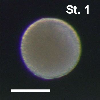
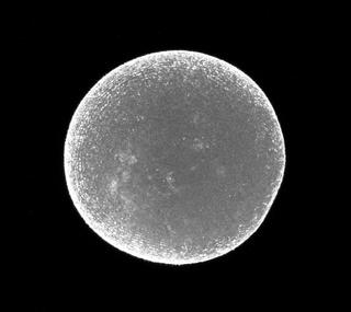
0.4hr | 2% | - | - | 接合体, 受精卵 |
|
|
|
| 0.8hr | 5% | - | - | - |
|
| ステージ | 受精後 経過時間 |
ふ化までの 時間経過(%) |
尾部の長さ(µm) | 尾部 / 体幹部 比 | 特徴 | リンク | 画像 (実体顕微鏡) |
画像 (3D構築) |
|---|---|---|---|---|---|---|---|---|
|
2 |
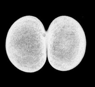
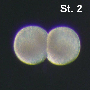
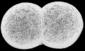
0.9hr | 6% | - | - | 2細胞胚 |
|
|
|
|
3 |
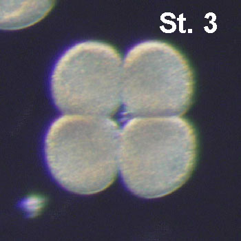
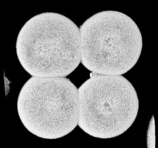
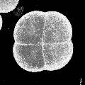
1.45hr | 8% | - | - | 4細胞胚 |
|
|
|
| 1.7hr | 10% | - | - | - |
|
|||
|
4 |
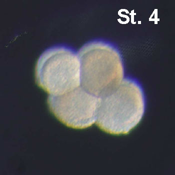
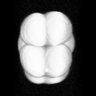
1.9hr | 11% | - | - | 8細胞胚 |
|
|
|
|
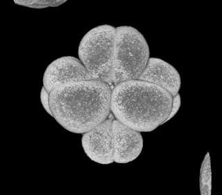
5a |
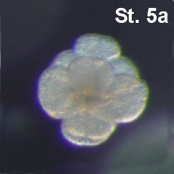
2.35hr | 13% | - | - | 16細胞胚の前期 |
|
|
|
|
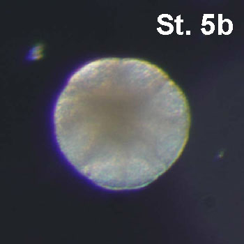
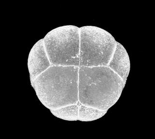
5b |
2.65hr | 15% | - | - | 16細胞胚の後期 |
|
|
|
|
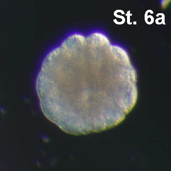
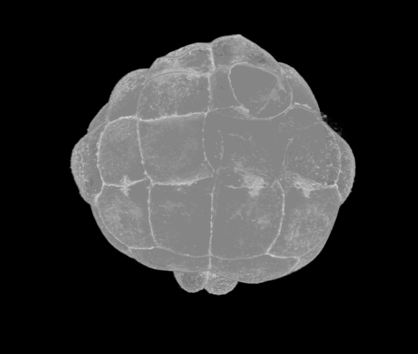
6a |
3hr | 17% | - | - | 32細胞胚の前期 |
|
|
|
|
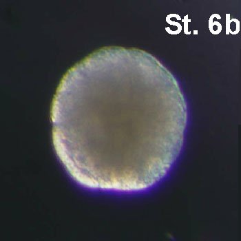
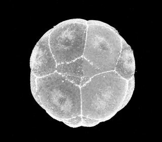
6b |
3.2hr | 18% | - | - | 32細胞胚の後期 |
|
|
|
|
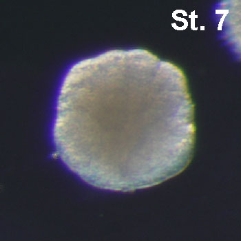
7 |
3.35hr | 19% | - | - | 44細胞胚。胚の植物半球が非常に丸みを帯びる。 |
|
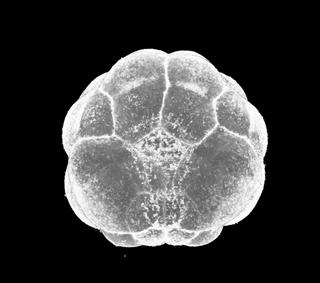
|
|
|
8 |
4hr | 23% | - | - | 64細胞胚。胚の植物半球が非常に丸みを帯びる。 |
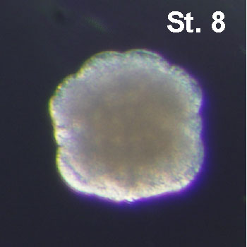
|
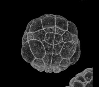
|
|
|
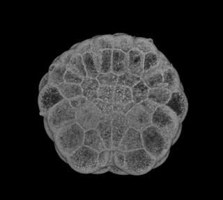
9 |
4.2hr | 24% | - | - | 76細胞胚。胚の植物半球は非常に平坦である。 |
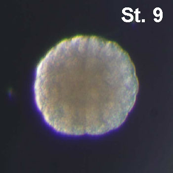
|
|
| ステージ | 受精後 経過時間 |
ふ化までの 時間経過(%) |
尾部の長さ(µm) | 尾部 / 体幹部 比 | 特徴 | リンク | 画像 (実体顕微鏡) |
画像 (3D構築) |
|---|---|---|---|---|---|---|---|---|
|
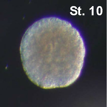
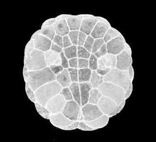
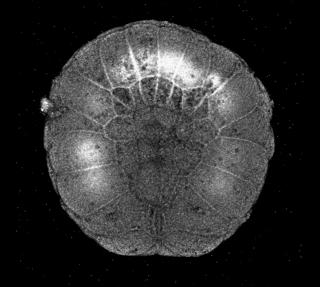
10 |
4.5hr | 26% | - | - | A7.1割球の頂端収縮を伴い、原腸陥入が開始する。 |
|
|
|
| 4.6hr | 27% | - | - | - |
|
|||
|
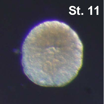
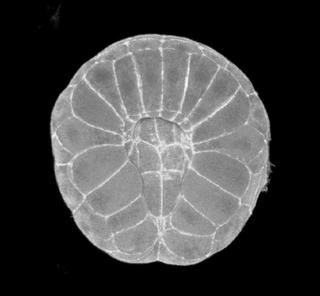
11 |
4.9hr | 28% | - | - | 脊索が陥入していく。胚の植物半球が蹄鉄のような形状になる。 |
|
|
|
|
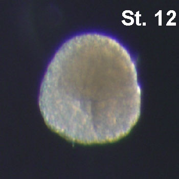
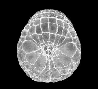
12 |
5.65hr | 32% | - | - | 神経板の割球が6列に配置される。原口はまだ胚の中央に位置し、開いたままである。 |
|
|
|
|
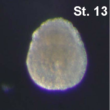
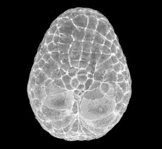
13 |
5.9hr | 34% | - | - | 原口は胚の後方に位置しており、ほとんど閉鎖している。胚は前後軸方向に伸長している。神経板は6列以上になり、A系列の神経割球列(I and II)がカーブし始める(神経管形成開始)。b6.5の大きな子孫細胞は胚の正中線付近に移動する。 |
|
|
| ステージ | 受精後 経過時間 |
ふ化までの 時間経過(%) |
尾部の長さ(µm) | 尾部 / 体幹部 比 | 特徴 | リンク | 画像 (実体顕微鏡) |
画像 (3D構築) |
|---|---|---|---|---|---|---|---|---|
|
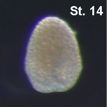
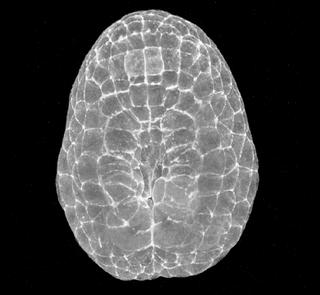
14 |
6.35hr | 36% | - | - | A系列の神経板がb6.5割球の娘細胞によって並ばせられ、溝を形成する。胚はひし形になる。溝は閉じ切っていない。 |
|
|
|
|
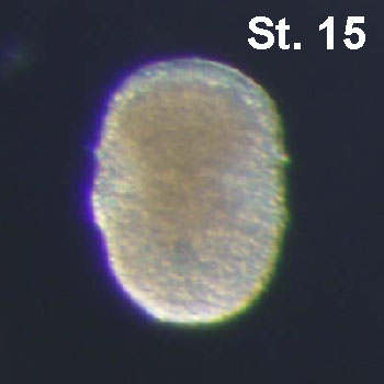
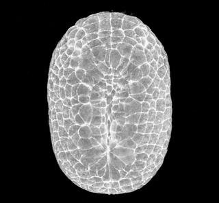
15 |
6.8hr | 39% | 83.0 | 1.0 | 神経管がほぼすべて形成される。胚は楕円形になる。A系列の神経板も溝を形成する。 |
|
|
|
|
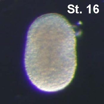
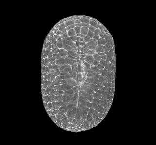
16 |
7.4hr | 42% | 84.2 | 1.0 | 神経管が後方の領域でも形成されはじめる。胚は前後軸方向に伸長していく。 |
|
|
| ステージ | 受精後 経過時間 |
ふ化までの 時間経過(%) |
尾部の長さ(µm) | 尾部 / 体幹部 比 | 特徴 | リンク | 画像 (実体顕微鏡) |
画像 (3D構築) |
|---|---|---|---|---|---|---|---|---|
|
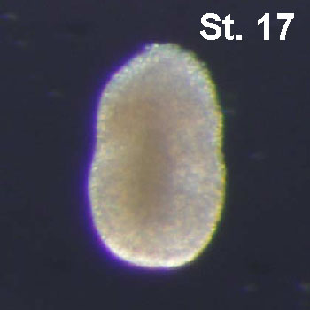
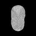
17 |
8.45hr | 48% | 87.8 | 1.0 | 体幹部と尾部が分離する最初の兆候があらわれる。尾部は曲がっておらず、体幹部と同程度の長さである。どの脊索細胞もインターカレーション(細胞挿入)が終了していない。 |
|
|
|
|
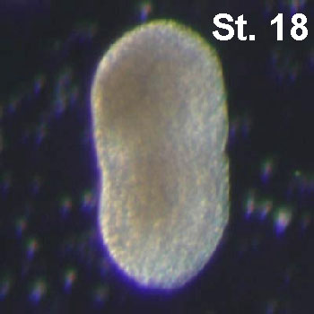
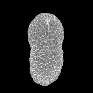
18 |
8.8hr | 50% | 111.9 | 1.1 | 尾部は体幹部からはっきりと区別できるようになる。尾部と体幹部はまだ同程度の長さである。A系列細胞の神経管形成によってあいた神経孔はまだ開いたままである。 |
|
|
|
|
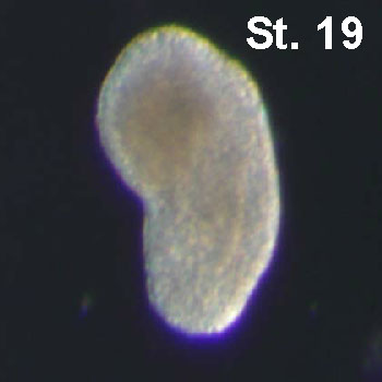
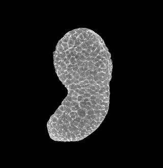
19 |
9.3hr | 53% | 120.3 | 1.2 | 尾部は腹側に約40°ほど曲がり、体幹部よりも少し長くなる。少数の脊索細胞がインターカレーションしはじめ、直線状に並んでいく。 |
|
|
|
|
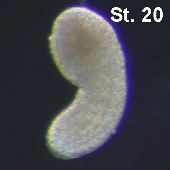
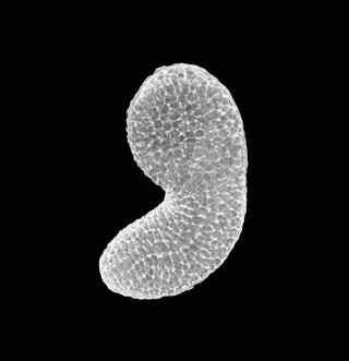
20 |
9.5hr | 54% | 146.7 | 1.3 | 神経孔が閉じ、神経管形成が完了する。尾部は約60°に曲がる。 |
|
|
|
|
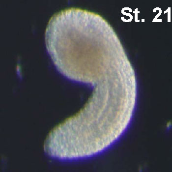
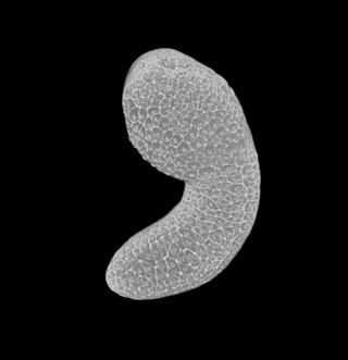
21 |
10.0hr | 57% | 180.7 | 1.6 | 尾部は体幹部の1.5倍の長さになり、腹側に約90°曲がっている。脊索細胞のインターカレーションが完了する。 |
|
|
|
|
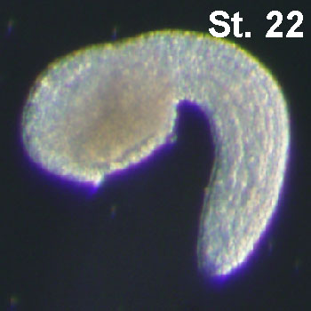
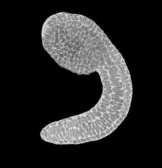
22 |
10.9hr | 62% | 221.2 | 1.9 | 胚の形が半円状をえがく。尾部は体幹部の2倍の長さになる。 |
|
|
|
|
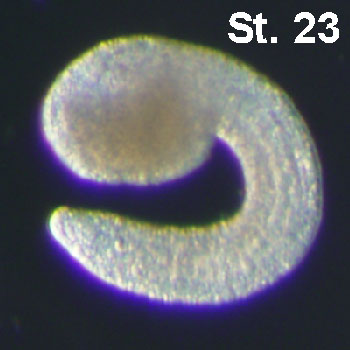
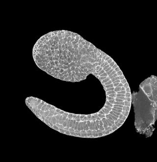
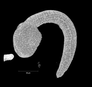
23 |
11.9hr | 68% | 255.1 | 2.1 | 平衡点の色素沈着が始まる。尾部は強く湾曲しており、尾部の先端が体幹部前方に近づいている。 |
|
|
|
| 12hr | 69% | - | - | - |
|
|||
|
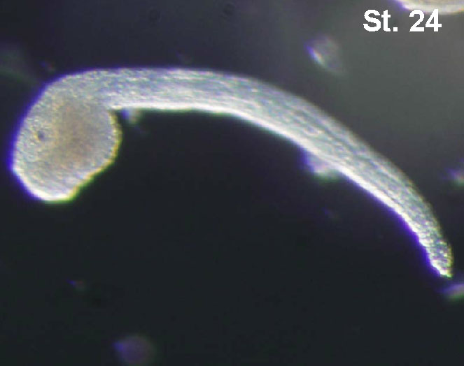
24 |
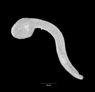
13.5hr | 77% | 442.6 | 3.4 | 脊索細胞の液胞化が始まる。胚の前端に付着器が生じ始める。尾部は曲がりが緩和して直線状になる。 |
|
|
|
|
25 |
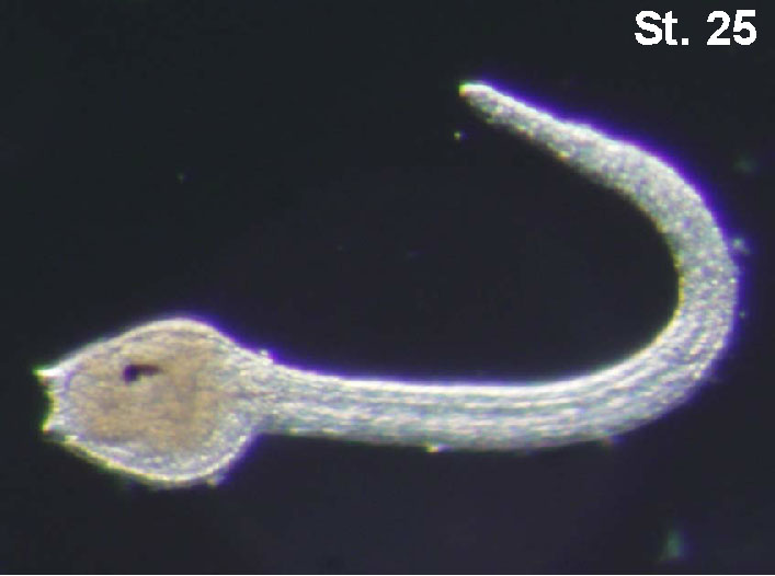
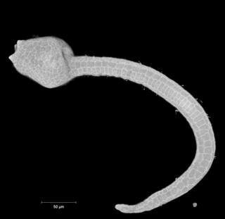
15.9hr | 91% | 558.6 | 3.9 | 眼点にメラニンが沈着する。すべての脊索細胞が液胞化する。尾部は背側に曲がりを生じる。 |
|
|
|
| 16.5hr | 94% | - | - | - |
|
|||
| 17.0hr | 97% | - | - | - |
|
| ステージ | 受精後 経過時間 |
ふ化までの 時間経過(%) |
尾部の長さ(µm) | 尾部 / 体幹部 比 | 特徴 | リンク | 画像 (実体顕微鏡) |
画像 (3D構築) |
|---|---|---|---|---|---|---|---|---|
|
26 |
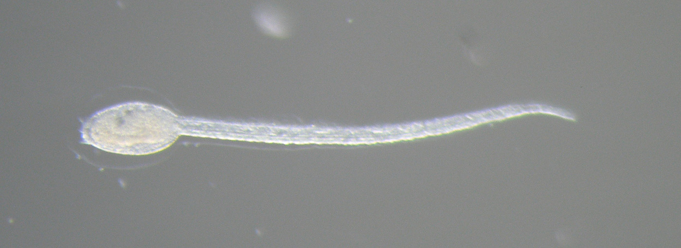
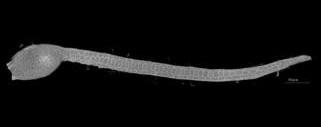
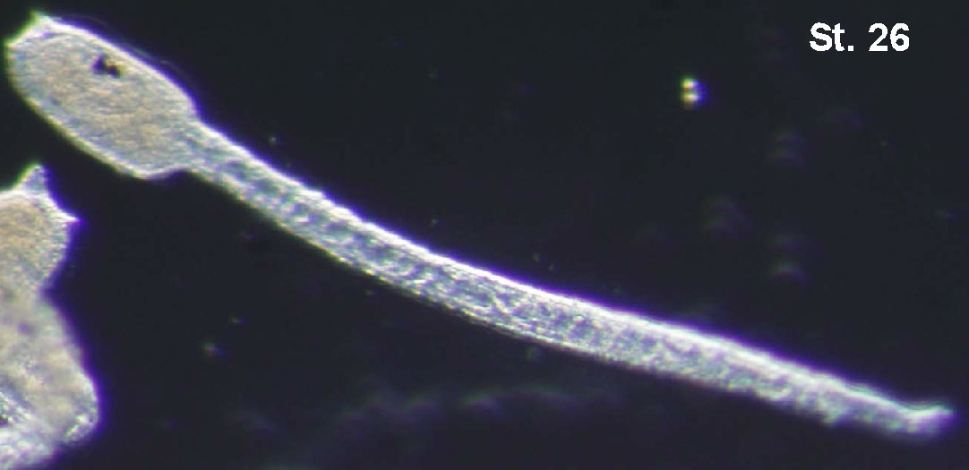
17.5hr | 100% | 661.6 | 4.2 | 卵殻をやぶってふ化する。体幹部は細長い長方形をしている。 |
|

|
VI. 幼生 Larva Period (St.26~30, 17.5~23*hpf**)
| ステージ | 受精後経過時間 | 特徴 | リンク | 画像 (実体顕微鏡) |
画像 (3D構築) |
|---|---|---|---|---|---|
| 17.5hpf | 卵殻をやぶってふ化する。体幹部は球状に近い形をしている。付着器の突起は未成熟でピラミッド型である。尾は不規則な動きをする。 |

|
|
|
|
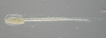
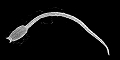
| 17.5-20 hpf | 体幹部は紡錘形をしている。尾は規則的な動きをして遊泳しはじめる。 |
|
|
|
|
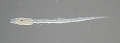
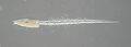
| 20-22hpf | 付着突起が伸長し、付着器の基底部が広がる。体幹部は角ばってくる。球状のテスト細胞、表皮感覚ニューロンの繊毛、口腔前葉が認識できるようになる。 |
|
|

|
|
|
|
|
|
|||
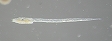
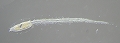
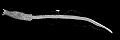
| 22-24hpf | ステージ28に比べ、体幹部はより長細い。体幹部と尾部の境界の輪郭は角ばっている。 |
|
|
|
|
|
|
|
|
VII. 付着 Adhesion period (St.31~32, 24~26hpf)
| ステージ | 受精後経過時間 | 特徴 | リンク | 画像 (実体顕微鏡) |
画像 (3D構築) |
|---|---|---|---|---|---|
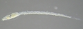
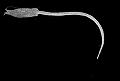
| 24 hpf | 付着突起は湾曲する。眼点と平衡点の残遺物が認識できる。 |
|
|
|
|
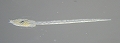
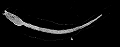
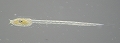
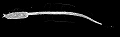
| 25-27 hpf |
|
|
|
||
|
|
|
|
VIII. 尾部退縮 Tail absorption period (St.31~33, 27~30hpf)
| ステージ | 受精後経過時間 | 特徴 | リンク | 画像 (実体顕微鏡) |
画像 (3D構築) |
|---|---|---|---|---|---|
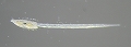
| 27 hpf | 尾部の吸収が始まる。体幹部と尾部の境界で尾が屈曲する。まだ眼点と平衡点の残遺物が認識できる。 |
|
|
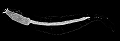
|
|
| 28 hpf | 尾部の半分が体幹部に吸収される。尾は収縮して厚くなる。まだ眼点と平衡点の残遺物が認識できる。 |
|
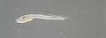
|
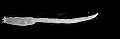
|
|
| 29 hpf | 尾部は完全に体幹部に吸収される。付着突起は識別できない。まだ眼点と平衡点の残遺物は認識できる。 |
|
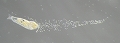
|
|
IX. 体軸回転 Body axis rotation period (St.34~36, 30~60hpf)
| ステージ | 受精後経過時間 | 特徴 | リンク | 画像 (実体顕微鏡) |
画像 (3D構築) |
|---|---|---|---|---|---|
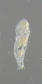
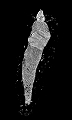
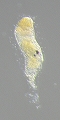
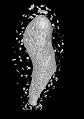
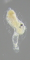
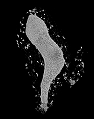
| 30-36 hpf | 体軸の回転が始まる(柄と内柱*のなす角度が0°を超える)。外側の被嚢とキューティクル層はもはや存在せず、一部被嚢の細胞は認識できる。また眼点と平衡点の残遺物も認識できる。 |
|
|
|
|
|
|
|
|
|||
|
|
|
|
|||
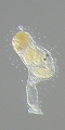
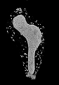
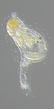
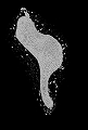
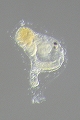
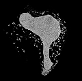
| 36-45 hpf | 体軸の回転が30°~60°ほどまで進行する。1対の鰓裂が認識できる。眼点と平衡点の残遺物も認識できる。 |
|
|
|
|
|
|
|
|
|||
|
|
|
|
|||
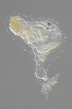
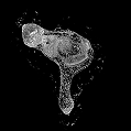
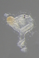
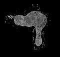
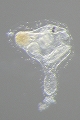
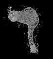
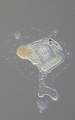
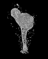
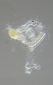
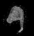
| 45-60 hpf (2dpf**) | 体軸の回転が80°~90°ほどまで進行する。2対の鰓裂が認識できる。眼点と平衡点の残遺物も認識できる。濾過摂食の活動や心臓の拍動が見られる。 |
|
|
|
|
|
|
|
|
|||
|
|
|
|
|||
|
|
|
|
|||
|
|
|
 |
| ステージ | 受精後経過時間 | 特徴 | リンク | 画像 (実体顕微鏡) |
画像 (3D構築) |
|---|---|---|---|---|---|
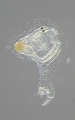
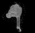
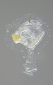
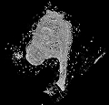
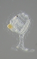
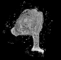
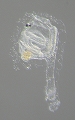
| 63-72 hpf (3dpf) | 体軸の回転が完了する。胃が膨張する。眼点と平衡点の残遺物が認識できる。 |
|
|
|
|
|
|
|
|
|||
|
|
|
|
|||
|
|
|
|
|||
|
38 前期 |
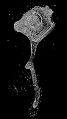
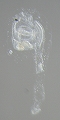
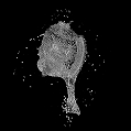
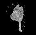
4 dpf | 幼生の尾部の残遺物が完全に吸収される。 |
|
|
|
|
|
||||
|
39 中期 |
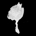
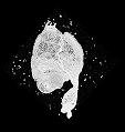
6 dpf | さらに鰓裂が開き始める。胃や腸管、神経が目立つようになる。 |
|
|
|
|
|